Author: admin
Ankle Sprain
Posted on August 15, 2018 by admin
An ankle sprain occurs when the ligaments that help hold the ankle bones together are forced beyond their normal range of motion. The ligaments on the outer side of the ankle are usually stretched, teared partially, or teared completely. Rolling, twisting, or turning your ankle in an awkward way can lead to an ankle sprain. It can be caused from walking or exercising on an uneven surface or from falling. Someone who has injured their ankle before or wears improper shoes increases their chance of a sprained ankle. It is also a very common sports injury, especially in soccer, tennis, and basketball because these sports involve jumping, cutting action, or twisting of the foot.
Depending on how severe the sprain is, the signs and symptoms include tenderness when the ankle is palpated, pain when weight is applied on the affected foot, swelling, bruising, limited range of motion, ankle instability, or popping sound heard at the time of injury.
Rest, Ice, Compression, and Elevation (R.I.C.E.) can be done to treat the sprained ankle for the first 2-3 days. Depending on the severity, sports tape or an ankle support brace can be used for stabilizing the ankle. In order to avoid an ankle sprain, one should warm up before exercising or playing sports, work on muscle strength and flexibility, and perform balancing exercises.
Stretching and Flexibility
Posted on August 7, 2018 by admin
As we get older our muscles become stiffer and we would start to notice our flexibility declining. The muscles will decrease in length if we don’t stretch our muscles. Shortened muscles could increase our risk for falling and one might find it difficult to perform activities that require flexibility, such as going up the stairs. In addition, using a shortened muscle for activity could also lead to muscle damage, strains, and joint pain.

Stretching can help us become more flexible and it is the key to preventing injury and disability. It can increase our range of motion and reduce joint and back pain. Moreover, it can decrease the risk of falling by improving our balance and can also improve our posture. Lastly, it can reduce the risk for muscle and joint injury.
It is important to warm up before we stretch the muscles. We can warm up by doing 2 to 5 minutes of dynamic stretching, such as lifting our knees and rolling our shoulders. This type of stretch involves moving a joint repeatedly through its available range of motion and does not involve holding a position. Static stretches can then be held for 10 to 30 seconds in order to regain flexibility and should be performed after a workout.
Herniated disk
Posted on July 27, 2018 by admin
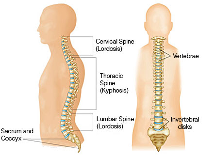
The spine consists of spinal disks and vertebrae and similar to a jelly donut, the disk has a softer “jelly” center. When the “jelly” pushes out through a tear in the tougher exterior, it is known as a herniated disk. Disk degeneration can cause disk herniation in the lower back or in the neck. The water content in the disks decrease as we get older and the flexibility of it decreases. Therefore, a minor strain, twist, and improper lifting can easily lead to a tear or rupture in the disk.
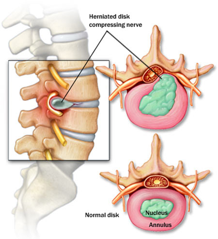
The signs and symptoms that someone will experience includes pain, numbness, tingling, or muscle weakness due to the irritation on the nerves. Pain can be felt in the thigh and calf when the herniated disk is in the lower back. Pain can be felt in the shoulder and arm when the herniated disk is in the neck.
Excess body weight and duties performed on the job that put extra stress on your lower back can increase the risk of disk herniation. A few things that you can do to prevent a herniated disk includes lifting heavy objects properly, strengthening trunk muscles, and maintaining a healthy weight.
Heel Spurs
Posted on July 18, 2018 by admin
Heel spurs are also known as calcaneal spurs or osteophytes. Someone with this condition would find a calcium deposit forming a bony protrusion along the plantar fascia. Running and jumping, especially on hard surfaces can wear down the heel and lead to heel spurs. If the membrane that covers the heel bone is torn or if the heel is bruised, it can cause heel spurs. Older adults, obesity, plantar fasciitis, and improper footwear can increase the chance of developing heel spurs.
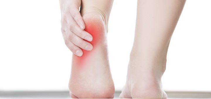
Heel spurs can lead to a sharp pain in the heel when he or she stands up, a dull ache in the heel, and heat radiating from the affected area. Tenderness, swelling, and inflammation can occur at the heel and a bone-like protrusion beneath the heel may also be seen. One can reduce pain and swelling by resting and applying ice to the affected area. Orthotics can also be worn to remove some of the pressure off the heel.
Myositis Ossificans
Posted on July 11, 2018 by admin
Myositis ossificans is when bone tissue forms within a muscle. It is usually found in the thigh muscles where the hamstring and quadricep muscles are found. Myositis ossificans can develop when a contusion (bruise), repetitive trauma, or strain occurs to a muscle. Calcification and bony formation can occur in the injured muscle when a repetitive trauma occurs before the injured area has been completely recovered. Therefore, it is important to rest the muscle after an injury. To prevent a repetitive trauma from happening, one can use protective padding and perform stretches to avoid muscle strain. Intensive stretching and massage should be avoided because it inhibits healing and increases bleeding into the muscle. Myositis ossificans can also occur if the inflammation and swelling of the muscle is ignored after an injury.
Moreover, myositis ossificans can lead to swelling, pain when exercising, limited range of motion, a weaker limb, and a hard bump. However, it can be treated by resting the muscle, strengthening the injured muscle and the muscles around it, and working on flexibility and light stretching. In addition, ultrasound can also help with the healing process.
Bursitis
Posted on July 4, 2018 by admin
Bursae are small, fluid-filled sacs that act as a cushion for the bones, tendons, and muscles around the joints. When they become inflamed, it is known as bursitis. Bursitis can occur in the shoulder, hip, elbow, and knee. Repetitive movements or positions that can apply pressure on the bursae, such as lifting something over your head repeatedly or kneeling on your knees for a long time can lead to bursitis. Inflammatory arthritis, an injury or a trauma can also cause bursitis. Someone who has bursitis will notice their joint becoming red and swollen. In addition, the joint will feel achy or stiff and they will feel pain when they try moving the joint or when they apply pressure to it.
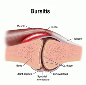
Older adults, overweight individuals, and individuals with rheumatoid arthritis have a higher risk of developing bursitis. However, one can reduce their risk by warming up and stretching before exercising, strengthening their muscles, lifting properly, carrying lighter loads, and taking breaks after performing repetitive movements. Rest, ice, and exercises can help relieve the pain. A corticosteroid drug can be injected into the bursa to reduce pain and inflammation. The pain will disappear within a few weeks if it is treated properly.
Osteoporosis
Posted on June 27, 2018 by admin
Osteoporosis is when your bones become thin and brittle. It is caused by a lack of bone strength or bone density. Similar to a sponge, your bones contain numerous holes which makes it fragile and can lead to hip, spine, or wrist fractures. This disease is usually found in older adults after age 60 and women are more likely to have osteoporosis than men. The risk of getting osteoporosis increases as we age and a woman who has gone through menopause has a greater risk. Other risk factors include a slender body, family history, people of European and Asian background, smoking, alcoholics, not getting enough weight-bearing exercises, and a lack of calcium and vitamin D intake.
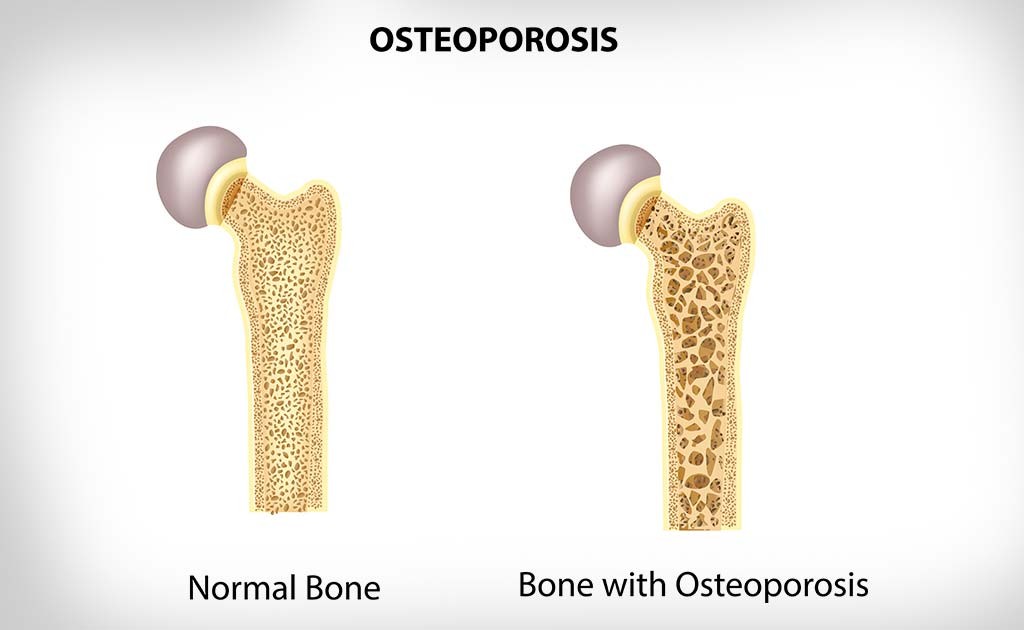
The first sign of osteoporosis is usually seen after a fall or bump where a fracture is found in their hip, spine, or wrist. They would later on experience back pain, notice their height has shrunk, and notice a curved backbone.
A bone density test can be completed to measure the thickness of your bones and the risk for a fracture. Calcium and vitamin D is essential to building strong, healthy bones. In addition, exercising and quitting smoking can help slow osteoporosis. In order to avoid bone fractures from occurring, it is important to get a bone density test and get osteoporosis treated early.
Lordosis
Posted on June 22, 2018 by admin
Lordosis is located in your neck and spine. Cervical lordosis is when there is an inward curve in the region of your neck. When there is an inward curve in your lower back, it is known as lumbar lordosis. It is more common for an individual to have lumbar lordosis than cervical lordosis. Obesity, osteoporosis, poor posture, spondylolisthesis, and kyphosis are a few causes of lordosis. Someone who does not have this condition would notice their ears are lined up over their hips, shoulders, and ankles.
Individuals who experience lordosis would have a noticeable curve that could be seen. In addition, they might experience discomfort, severe back pain and/or tense muscles around the curve. Lordosis could restrict their movements and create a space in between their neck or lower back and the floor when they are laying down.
Physical therapy can help with strengthening, and increase your flexibility and range of motion for your neck and spine. Exercising, stretching, using extra pillows when you go to sleep to support your spine, and wearing a neck immobilizer or thoracic spine brace are all possible treatment options for lordosis.
Kyphosis
Posted on June 18, 2018 by admin
A healthy spine is made up of vertebrae that look like cylinders put on top of one another to form a column. Kyphosis is when the vertebrae in the upper back is more wedge shaped, which leads to an excessive, forward rounding of the back. It can appear in infants or teens, but it is most commonly found in older women.
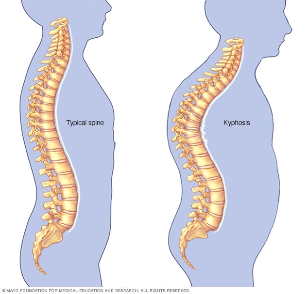
In addition to an abnormal curvature of the spine, someone who has mild kyphosis can experience back pain and stiffness. Fractures, disk degeneration, birth defects, osteoporosis can all lead to an abnormal vertebrae. Kyphosis is linked to weak back muscles and an individual can have difficulty walking, driving, and looking up. Pain can also be felt when the individual lies down and there can be problems with breathing and digestion if the situation is severe. Depending on the individual’s age and the cause, the treatment can vary for each individual.
Ankle Rehabilitation
Posted on June 6, 2018 by admin
Ankle sprains are a common injury that can lead to severe pain and weakness. We can work on various rehabilitation exercises to help our ankle heal and prevent future injuries.
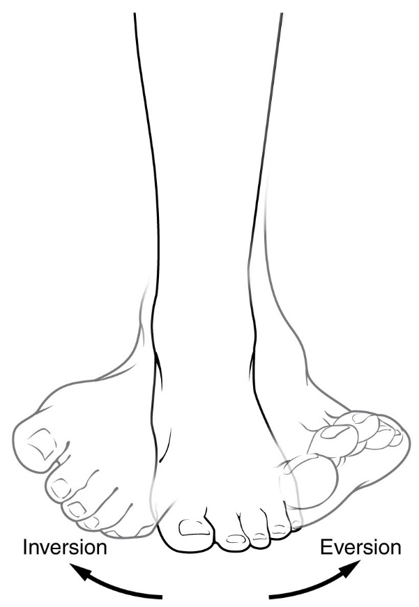
After the injury has occurred, it is important to perform partial weight bearing to prevent muscle atrophy. When you are able to walk again without limping, you can work on full weight bearing. You should begin with range of motion exercises, such as plantar flexion, dorsiflexion, inversion, and eversion. You should not feel pain when you perform these ankle movements. You can then practice writing the alphabet with your toe and work on towel assisted stretches with the same ankle movements.

Next, you can do strengthening exercises, such as performing the ankle movements against the wall or using a rubber tubing looped around your feet for resistance. After, you can move on to balance exercises on a wobble board by doing a double leg stand with eyes opened and then closed. Then, standing on your injured foot with eyes opened and then closed.
By following these rehabilitation steps, you can help strengthen the ankle and prevent an injury from occurring again.