22 September 2026 · Chiropractic
Shoulder injuries: rotator cuff, sprains and calcific tendinitis
Shoulder pain shows up after a fall on the ice, a hard weekend of painting, or months of desk work with no clear “injury day.” The shoulder is mobile by design — a shallow ball-and-socket that trades stability for reach — so tendons, ligaments and the joint capsule take a lot of load. Most problems improve with sensible load management, time, and a rehab plan. This article walks through three patterns we see often at Rupert Health Centre in East Vancouver (Collingwood / Kingsway area): rotator cuff injuries, shoulder girdle sprain and strain, and calcific tendinitis.
General education only. It is not a diagnosis or a personal treatment plan. Sudden deformity, inability to lift the arm after trauma, fever with a hot swollen joint, or progressive neurological loss needs urgent medical care.
Anatomy overview: what has to work together
Three bones form the shoulder girdle you feel under the skin: the clavicle (collar bone), the scapula (shoulder blade), and the humerus (upper arm). The ball of the humerus sits in the glenoid socket of the scapula. Above that sits the acromion — a bony roof that forms a tunnel (the subacromial space) where tendons pass. Ligaments stitch bone to bone (for example at the acromioclavicular or “AC” joint). Muscles and tendons move the arm and keep the ball centred in the socket.
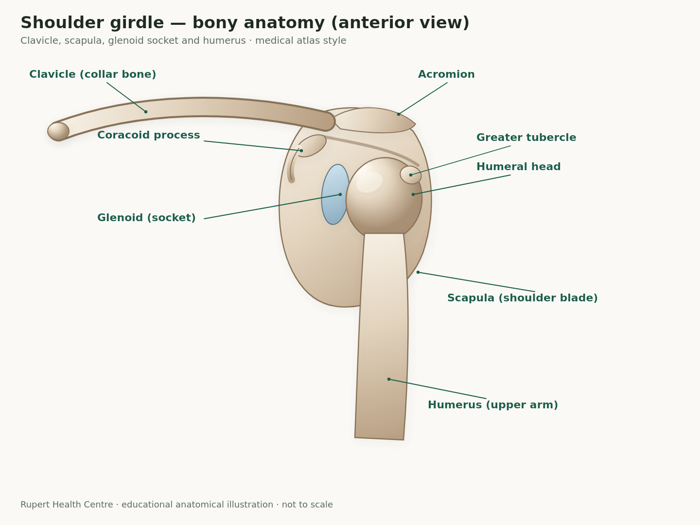
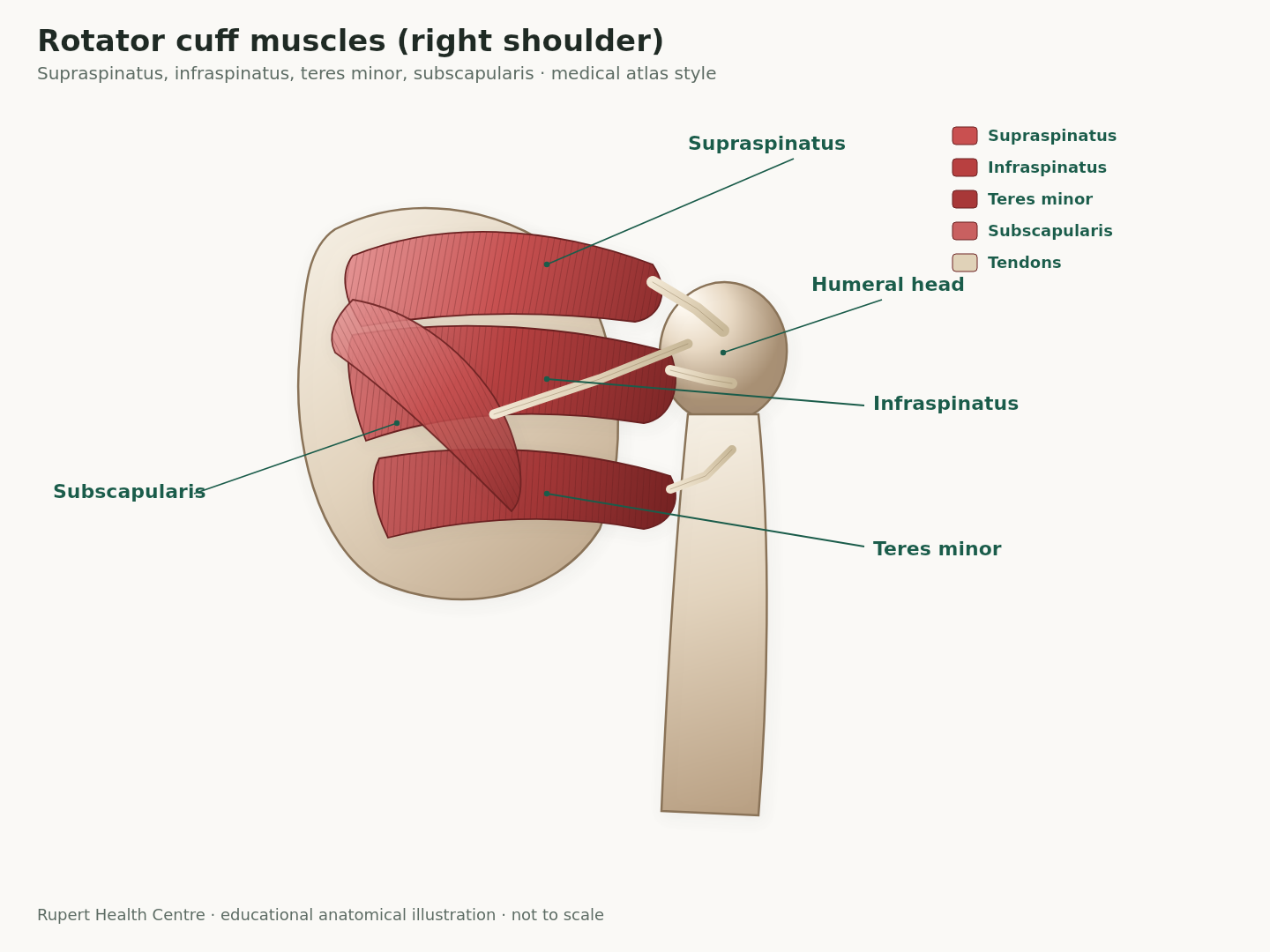
The rotator cuff is a group of four muscles — subscapularis, supraspinatus, infraspinatus and teres minor — whose tendons blend around the humeral head. Together they rotate the arm and, just as importantly, keep the ball from riding up into the roof of the shoulder when you lift. When cuff tendons are irritated or partially torn, people often feel a deep ache, night pain, and weakness with overhead reach — patterns described in patient education from Mayo Clinic and the American Academy of Orthopaedic Surgeons (AAOS OrthoInfo).
Rotator cuff injuries: tendinopathy and tears
What goes wrong
Rotator cuff problems sit on a spectrum. Early on, tendons can become irritated and thickened (tendinopathy / tendinitis language is still common in clinics). Over time, fibres can fray into a partial-thickness tear or, less often, a full-thickness tear. AAOS OrthoInfo notes two broad pathways: an acute tear after a fall or sudden load, and degenerative wear that accumulates with age and repetitive overhead work. Painters, carpenters, throwers and racquet athletes load these tendons often; risk also rises after about age 50.
Common signs
- Dull ache deep in the shoulder, often worse at night or when lying on that side.
- Pain or weakness reaching overhead, combing hair, or putting on a coat.
- Difficulty lifting or rotating the arm away from the body.
- After a clear injury: a pop, sudden pain, and immediate weakness — that pattern deserves prompt assessment.
When imaging helps
History and a careful exam come first. X-rays do not show the cuff tendons themselves, but they can reveal bone spurs, arthritis, or calcium deposits. Ultrasound or MRI is more useful when weakness is clear, trauma was significant, recovery has stalled, or a surgeon needs a map of tear size and tendon quality. Mayo Clinic’s guidance on rotator cuff injury emphasises that many people improve with activity modification, ice, medicines that are safe for them, and structured physical therapy — surgery is reserved for selected cases where pain and function stay limited.
Asymptomatic cuff changes also show up on scans in people without pain, especially with age. Imaging answers a clinical question; it does not automatically dictate surgery.
Shoulder sprain vs strain
Patients (and busy clinics) often use “sprain” and “strain” interchangeably. The structures differ:
- Sprain — stretch or tear of a ligament (bone-to-bone). Around the shoulder this often means the AC joint after a fall onto the point of the shoulder, or capsular/ligament strain after a wrenching motion. Bruising, local tenderness over a joint line, and pain with cross-body or overhead load are common.
- Strain — stretch or tear of a muscle or tendon. Cuff strains, deltoid strains, and pectoral strains after a heavy press or awkward lift fit here. Pain with resisted contraction of that muscle is a useful clue.
Grade language (mild stretch → partial fibre tear → more complete disruption) is less important on day one than calming swelling, protecting the joint from re-injury, and restoring motion without forcing painful end-range. After a car collision, shoulder girdle sprains and strains often travel with neck symptoms; we can discuss ICBC billing for chiropractic and active rehab on a separate visit if that applies (see our ICBC FAQ).
Calcific tendinitis (calcific tendonitis)
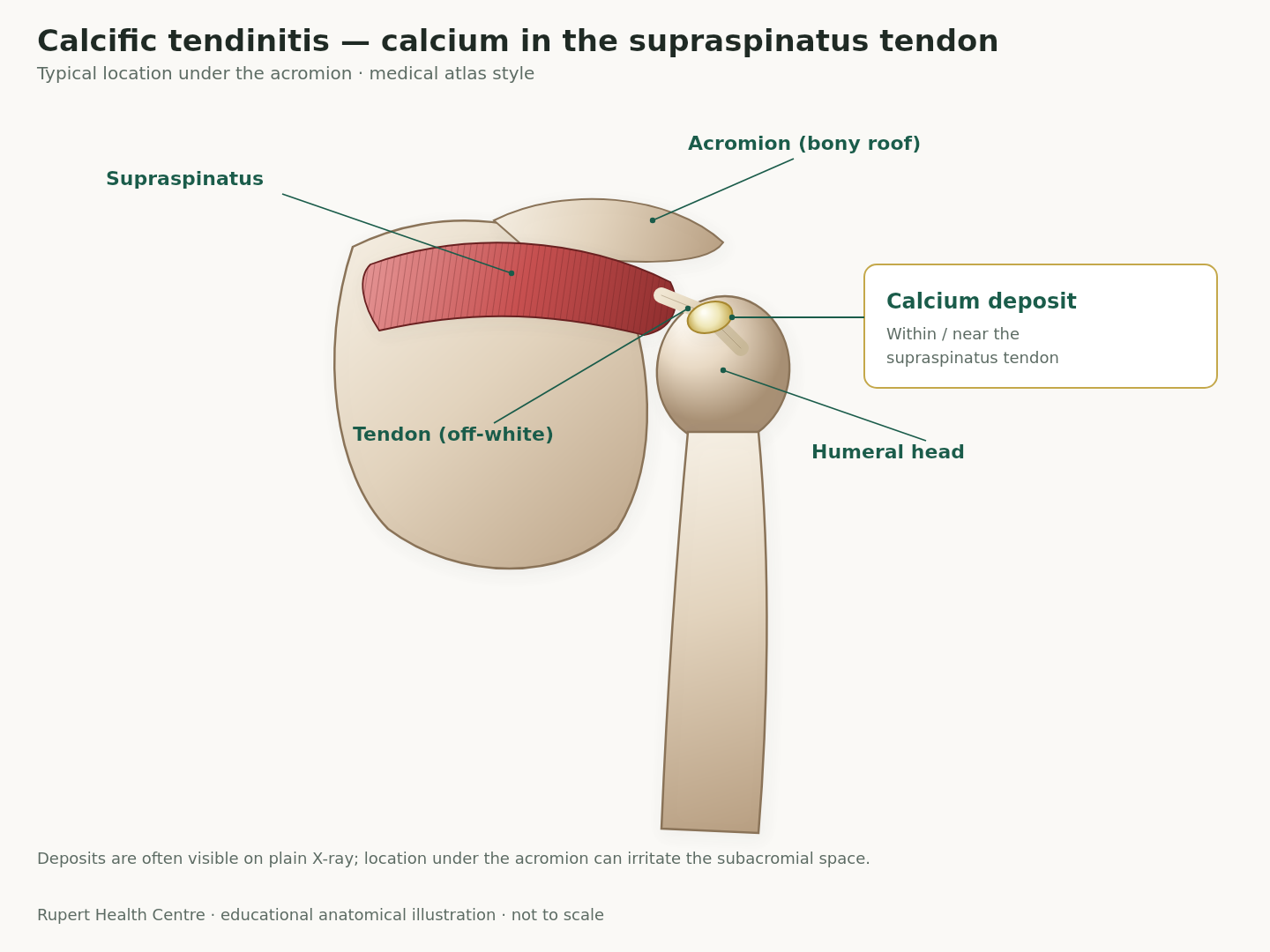
Calcific tendinitis is the build-up of calcium deposits inside a rotator cuff tendon — most often the supraspinatus. AAOS OrthoInfo explains that deposits can contribute to inflammation and mechanical irritation under the acromion. It is more common in adults roughly 30–60 years old and slightly more common in women; the exact cause is still unclear and may relate to tendon overuse or local tissue environment rather than diet “calcium excess.”
Quiet phase vs flare (resorptive) phase
Deposits can sit quietly for long periods with little pain. During a resorptive or inflammatory phase, the body begins breaking the deposit down. That phase can feel dramatic — severe night pain, marked loss of motion, and a shoulder that refuses to tolerate even light reach — even though the long-term outlook is often favourable as the deposit shrinks. X-rays help confirm calcium; ultrasound or MRI can clarify tendon quality and bursal inflammation if the picture is mixed.
Typical course and care
Many flares settle with relative rest from aggravating overhead work, pain control that is safe for you, and guided rehab to restore motion once the sharp phase eases. Persistent deposits or stubborn pain may lead your family doctor or orthopaedic clinician to discuss injection options or, less often, procedures to remove calcium. Our role in clinic is usually to keep the scapula and thoracic spine moving well, calm related soft tissue, and progress loading so the cuff and deltoid share work again — not to “dissolve calcium” with one adjustment.
What chiropractic and manual care typically address
At Rupert Health Centre (chiropractic and massage), a shoulder visit usually starts with mechanism, night pain, weakness testing, and screening for referral from the neck. Treatment is rarely “shoulder crack only.” We often combine:
- Joint mobilization or carefully selected manipulation of the thoracic spine, ribs, and sometimes the glenohumeral or AC joints when stiffness is limiting reach.
- Soft-tissue work to the cuff, scapular stabilizers, and upper traps / pecs that guard after pain.
- Progressive rehab: isometrics early when motion is irritable, then external-rotation and scapular endurance work, then return to lifting or sport-specific drills.
- Massage therapy when muscle guarding and desk-related tightness dominate the picture.
A Cochrane review of manual therapy and exercise for rotator cuff disease found that, in the one high-quality trial matching common clinical practice against a placebo ultrasound control, differences in pain and function at follow-up were small and not clearly clinically important — while mild short-term soreness after treatment was more common than with placebo. In plain terms: hands-on care plus exercise is a reasonable, guideline-aligned option people use alongside activity modification, but it is not a magic fix, and expectations should stay honest. Exercise-based rehab remains a cornerstone of non-surgical care in OrthoInfo and Mayo guidance.
Red flags — when to seek urgent care
- Obvious deformity, or suspicion of dislocation or fracture after a fall or collision — emergency / urgent orthopaedic assessment.
- Sudden inability to lift the arm after trauma with clear weakness — do not wait weeks hoping it “loosens up.”
- Hot, red, swollen joint with fever or feeling systemically unwell — possible infection; seek medical care urgently.
- Progressive numbness, hand weakness, or symptoms that sound more like a neck nerve root problem with cord-type features — medical evaluation.
- Unexplained weight loss, cancer history, or night pain that is relentless and unrelated to position — prompt medical review.
Book a clinic assessment sooner (non-emergency) if pain is stalling work or sleep beyond a couple of weeks, strength is dropping, or you are stuck in a flare of known calcific tendinitis and cannot progress home exercises.
Practical self-care (conservative and evidence-aligned)
- Relative rest, not total shutdown. Avoid repeated painful overhead presses and heavy reaching for a short window; keep the elbow and hand moving, and walk as usual.
- Short ice or heat trials. Ice after aggravating activity helps some people; heat before gentle mobility helps others. Use what settles symptoms.
- Sleep position. Try not to lie directly on the sore side; a pillow supporting the arm in slight forward elevation can reduce night ache.
- Desk setup. Raise the monitor, keep elbows near the body, and take micro-breaks — desk-tight shoulders often feed cuff irritation (related: massage for desk-tight shoulders).
- Graded loading. When sharp pain settles, light external-rotation bands and scapular rows usually beat endless passive stretching alone.
- Medicines. Over-the-counter options help some adults if safe for stomach, kidney and heart history — check with a pharmacist or family doctor.
How this fits at Rupert Health Centre
We assess shoulders in the context of the neck, mid-back and work or sport demands. Care plans stay short on mystery and long on homework. Collision-related shoulder injuries can be billed through ICBC where appropriate; that paperwork is secondary to getting a clear diagnosis pathway and a rehab ladder you can follow between visits.
Related: Shoulder labrum tears: SLAP, Bankart, and what they mean — companion guide to the glenoid labrum, instability patterns and how they differ from cuff problems.
Related: Frozen shoulder (adhesive capsulitis): stages, causes and what helps — why the joint capsule stiffens, the three stages, and how it differs from a rotator cuff problem.
References
- American Academy of Orthopaedic Surgeons (OrthoInfo). Rotator Cuff Tears. https://www.orthoinfo.org/diseases--conditions/rotator-cuff-tears/
- American Academy of Orthopaedic Surgeons (OrthoInfo). Calcific Tendinitis of the Shoulder. https://www.orthoinfo.org/diseases--conditions/calcific-tendinitis-of-the-shoulder/
- Mayo Clinic. Rotator cuff injury — Symptoms and causes. https://www.mayoclinic.org/diseases-conditions/rotator-cuff-injury/symptoms-causes/syc-20350225
- Mayo Clinic. Rotator cuff injury — Diagnosis and treatment. https://www.mayoclinic.org/diseases-conditions/rotator-cuff-injury/diagnosis-treatment/drc-20350231
- Page MJ, et al. Manual therapy and exercise for rotator cuff disease. Cochrane Database Syst Rev. 2016;(6):CD012224. https://www.cochrane.org/evidence/CD012224_manual-therapy-and-exercise-rotator-cuff-disease
← Back to all posts · Book an assessment · 604.435.2285 · 2955 Kingsway